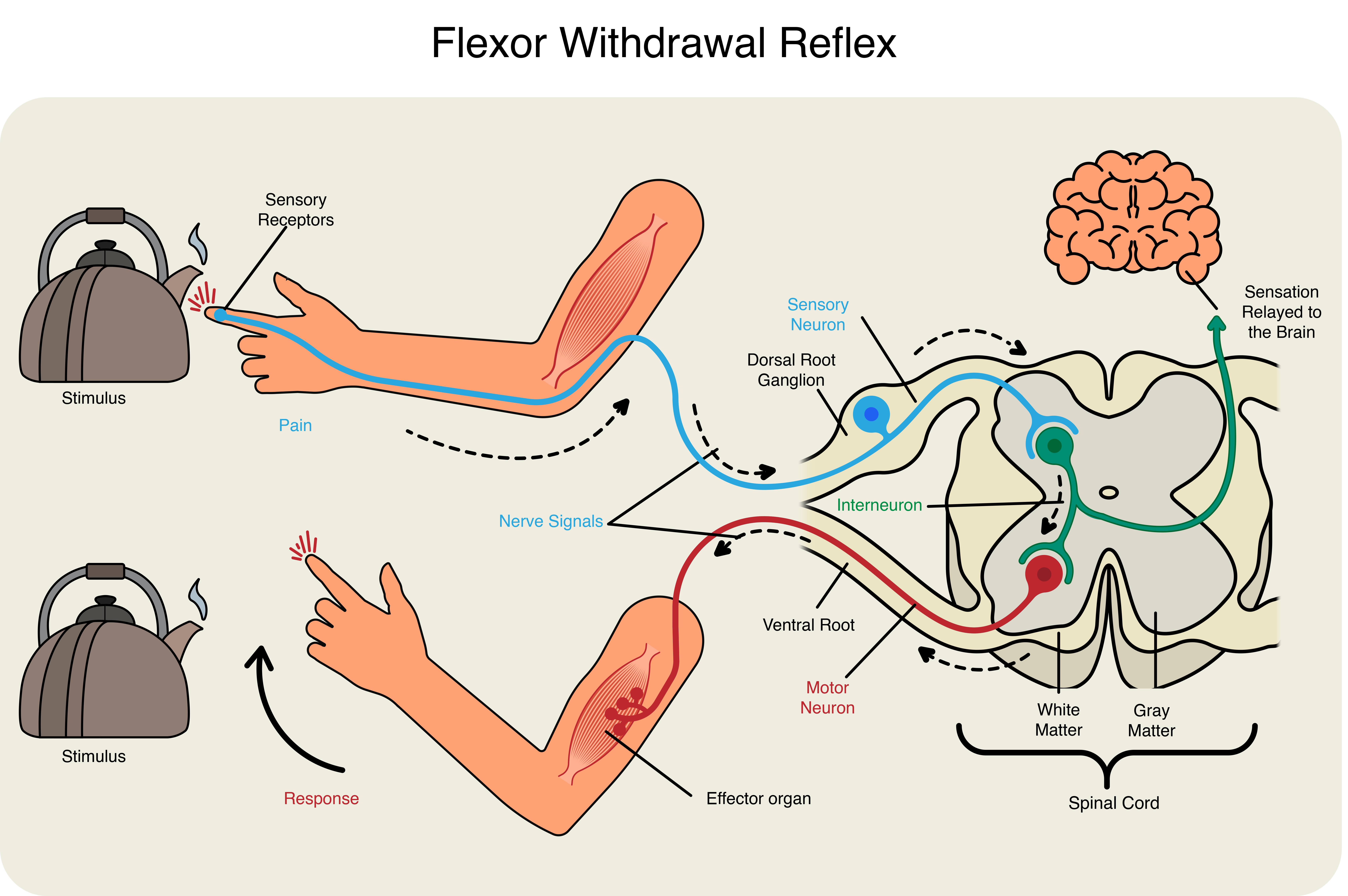
Reflexes can be spinal or cranial, depending on the nerves and central components that are involved. The body uses both spinal and cranial reflexes to rapidly respond to important stimuli. All reflex arcs include five basic components; (1) a receptor, (2) a sensory neuron, (3) an integration center, (4) a motor neuron, and (5) an effector. The effector may be a skeletal muscle, as is the case in somatic reflexes. However, in autonomic (or visceral) reflexes, the effector will be cardiac muscle, smooth muscle, or a gland.
Somatic spinal reflexes utilize motor neurons of the ventral horn of the spinal cord to activate skeletal muscles. The simplest example of this type of reflex is the stretch reflex. In this reflex, when a skeletal muscle is stretched, a muscle spindle receptor is activated. The sensory neuron associated with the muscle spindle synapses directly with the motor neuron in the ventral horn, allowing for an incredibly fast response called a monosynaptic reflex. This reflex helps maintain muscles at a constant length, and is the reason your head jerks back up after drooping when you begin to fall asleep sitting up. Another common example of this reflex is the knee jerk that is elicited by a rubber hammer struck against the patellar ligament in a physical exam.
Intrinsic and learned reflexes represent distinct categories of automatic, involuntary responses within the nervous system. Intrinsic reflexes, also known as innate or inborn reflexes, are hardwired and genetically predetermined. These reflexes are typically present from birth and do not require prior experience or learning. Examples of intrinsic reflexes include the knee-jerk reflex, where the tapping of the patellar tendon causes a rapid contraction of the quadriceps muscle and extension of the leg, and the withdrawal reflex, wherein the sudden touch of a hot object prompts the automatic removal of the affected body part.
On the other hand, learned reflexes, also referred to as acquired or conditioned reflexes, result from experience and training. These reflexes develop through repeated exposure to certain stimuli, leading to a conditioned response. A classic example is Pavlov’s experiment with dogs, where the ringing of a bell (neutral stimulus) became associated with the presentation of food, eventually causing the dogs to salivate (learned response) at the sound of the bell alone. Unlike intrinsic reflexes, learned reflexes involve a cognitive component and are acquired through the process of conditioning, demonstrating the adaptability of the nervous system based on environmental interactions and experiences.
Ipsilateral and contralateral reflexes refer to the specific sides of the body involved in a reflex arc. In ipsilateral reflexes, the sensory input and motor output occur on the same side of the body. An example of this is the withdrawal reflex, where the stimulus (such as touching a hot surface) causes a reflexive response, leading to the withdrawal of the affected limb on the same side without involving the opposite side. Contralateral reflexes, on the other hand, involve the sensory input on one side of the body and a motor output on the opposite side. An example is the crossed-extensor reflex, often observed in response to a painful stimulus. In this reflex, withdrawal of one limb on the side of the stimulus occurs (ipsilateral response), while the opposite limb on the other side extends to provide support (contralateral response). These reflex distinctions highlight the intricacies of neural pathways that govern motor responses and demonstrate the adaptive nature of the nervous system in coordinating complex movements.
A different somatic spinal nerve reflex involves the response to pain, like when you touch a hot stove and, in response, withdrawal your arm typically before you have even registered the pain in your hand. This reflex is called the flexor withdrawal reflex, and it stimulates the withdrawal of the arm through a connection in the spinal cord that leads to contraction of the biceps brachii (Figure 20.1). Unlike the stretch reflex, the flexor withdrawal reflex is polysynaptic and requires 2 spinal cord synapses to activate the motor neuron. As you withdraw your hand from the stove, you do not want to slow that reflex down. As the biceps brachii contracts, the antagonistic triceps brachii that had been activated to extend the arm toward the stove now needs to relax. Because the neuromuscular junction is strictly excitatory, the biceps will contract when the motor nerve is active. Skeletal muscles do not actively relax. Instead the motor neuron needs to “quiet down,” or be inhibited. In the hot-stove withdrawal reflex, this occurs through an interneuron in the spinal cord. The interneuron’s cell body is located in the dorsal horn of the spinal cord. The interneuron receives a synapse from the axon of the sensory neuron that detects that the hand is being burned. In response to this stimulation from the sensory neuron, the interneuron inhibits the motor neuron that controls the triceps brachii, in what is known as reciprocal inhibition. This is done by releasing a neurotransmitter or other signal that hyperpolarizes the motor neuron connected to the triceps brachii, making it less likely to initiate an action potential. With this motor neuron being inhibited, the triceps brachii relaxes. Without the antagonistic contraction, withdrawal from the hot stove is faster and keeps further tissue damage from occurring.

The flexor withdrawal reflex is also at play when you step on a painful stimulus, like a tack or a child’s Lego®. The nociceptors that are activated by the painful stimulus activate the motor neurons responsible for contraction of the tibialis anterior muscle. This causes dorsiflexion of the foot. An inhibitory interneuron, activated by a collateral branch of the nociceptor fiber, will inhibit the motor neurons of the gastrocnemius and soleus muscles to cancel plantar flexion. An important difference in this reflex is that plantar flexion is most likely in progress as the foot is pressing down onto the tack. Contraction of the tibialis anterior is not the most important aspect of the reflex, as continuation of plantar flexion will result in further damage from stepping onto the tack. While all this is happening in one lower limb, a contralateral response will be stimulated to help you catch your balance with the other. This is called the crossed extensor reflex.
In the cross extensor reflex, the same painful stimulus that initiates the flexor withdrawal reflex simultaneously initiates extension of the opposite limb. In the case of stepping on a painful object and pulling your foot away, the cross extensor reflex activates the contralateral quads and gastrocnemius and soleus to extend the leg while plantar flexing the ankle to shift body weight.
All of the somatic spinal nerve reflexes involved so far involve reciprocal inhibition. In each case, a prime mover is stimulated and its antagonist is inhibited. However, in the golgi tendon reflex, the prime mover is inhibited its antagonist is stimulated. This is termed reciprocal activation. In the tendon reflex, prolonged or particularly forceful stretching of the muscle and its tendon trigger the relaxation of the muscle to prevent tearing through the activation of a special receptor, the golgi tendon organ. At the same time, the antagonist muscle is activated to help return the affected muscle and its tendon to their resting lengths.
definitionan automatic, patterned response to a stimulus